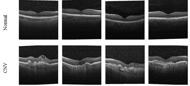
Eye is one of the primary sensory organs, responsible to provide the necessary visual information to the brain. Any abnormality in eye cause mild to severe vision issues and hence appropriate detection and treatment is preferred. Clinical level examination of the eye is commonly performed using image examination procedures and this work considered the Optical Coherence Tomography (OCT) for the study. In the present study, the proposed system aims to develop a tool to classify the OCT-images into normal and abnormal class using the traditional deep-learning (DL) and machine-learning (ML) features.
Methods
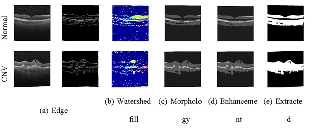
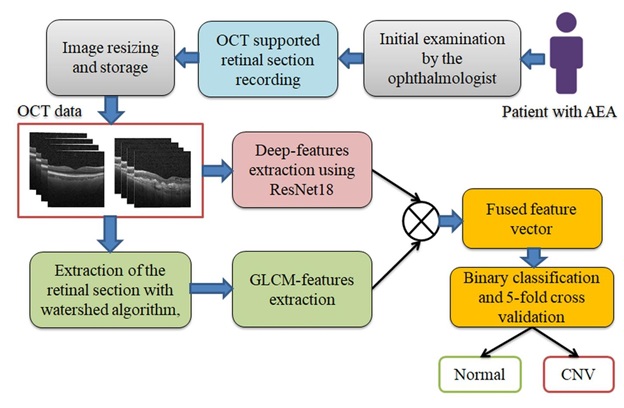
This tool consists the following stages; OCT-image collection and resizing based on the chosen DL scheme, image features extraction using DL and ML methods, DL feature reduction using 50% dropout and serially fusing the deep- and machine-features to generate hybrid-features, and binary classification using 5-fold cross-validation.
Results
The developed tool’s merit is verified using; (i) deep-features, (ii) machine-features, and (iii) hybrid-features. The merit of the developed scheme is verified using different binary classifiers and the overall quality metric is considered to verify the tool’s performance.
Conclusions
The study confirms that the hybrid-features based detection provides 97.6% accuracy, when Random Forest classifier is employed, which verifies the tool’s merit on the chosen OCT database. In the future, this tool can be considered to detect the common AEAs, like AMD and the DME.
Mārtiņš, A. (2025). Hybrid Image Features Supported Normal/Abnormal Retinal Optical Coherence Tomography Image Classification. International Journal of Clinical Medical Research, 3(1), 46. doi:10.61466/ijcmr3010001
ACS Style
Mārtiņš, A. Hybrid Image Features Supported Normal/Abnormal Retinal Optical Coherence Tomography Image Classification. International Journal of Clinical Medical Research, 2025, 3, 46. doi:10.61466/ijcmr3010001
AMA Style
Mārtiņš A.. Hybrid Image Features Supported Normal/Abnormal Retinal Optical Coherence Tomography Image Classification. International Journal of Clinical Medical Research; 2025, 3(1):46. doi:10.61466/ijcmr3010001
Chicago/Turabian Style
Mārtiņš, Aleksandrs 2025. "Hybrid Image Features Supported Normal/Abnormal Retinal Optical Coherence Tomography Image Classification" International Journal of Clinical Medical Research 3, no.1:46. doi:10.61466/ijcmr3010001
APA style
Mārtiņš, A. (2025). Hybrid Image Features Supported Normal/Abnormal Retinal Optical Coherence Tomography Image Classification. International Journal of Clinical Medical Research, 3(1), 46. doi:10.61466/ijcmr3010001
Article Metrics
Article Access Statistics
References
Zetterberg, M. (2016). Age-related eye disease and gender. Maturitas, 83, 19-26.
Chuck, R. S., Dunn, S. P., Flaxel, C. J., Gedde, S. J., Mah, F. S., Miller, K. M.,... & Musch, D. C. (2021). Comprehensive adult medical eye evaluation preferred practice pattern®. Ophthalmology, 128(1), P1-P29.
Ardeljan, D., & Chan, C. C. (2013). Aging is not a disease: distinguishing age-related macular degeneration from aging. Progress in retinal and eye research, 37, 68-89.
Bailey, S. T., Thaware, O., Wang, J., Hagag, A. M., Zhang, X., Flaxel, C. J.,... & Jia, Y. (2019). Detection of nonexudative choroidal neovascularization and progression to exudative choroidal neovascularization using OCT angiography. Ophthalmology Retina, 3(8), 629-636.
de Carlo, T. E., Bonini Filho, M. A., Chin, A. T., Adhi, M., Ferrara, D., Baumal, C. R.,... & Waheed, N. K. (2015). Spectral-domain optical coherence tomography angiography of choroidal neovascularization. Ophthalmology, 122(6), 1228-1238.
Bartlett, H., & Eperjesi, F. (2007). Use of fundus imaging in quantification of age-related macular change. Survey of ophthalmology, 52(6), 655-671.
Faridi, A., Jia, Y., Gao, S. S., Huang, D., Bhavsar, K. V., Wilson, D. J.,... & Bailey, S. T. (2017). Sensitivity and specificity of OCT angiography to detect choroidal neovascularization. Ophthalmology retina, 1(4), 294-303.
Umer, M. J., Sharif, M., Raza, M., & Kadry, S. (2023). A deep feature fusion and selection‐based retinal eye disease detection from OCT images. Expert Systems, e13232.
Yadav, S. S., & Jadhav, S. M. (2019). Deep convolutional neural network based medical image classification for disease diagnosis. Journal of Big data, 6(1), 1-18.
Talcott, K. E., Valentim, C. C., Perkins, S. W., Ren, H., Manivannan, N., Zhang, Q.,... & Singh, R. P. (2024). Automated detection of abnormal optical coherence tomography b-scans using a deep learning artificial intelligence neural network platform. International Ophthalmology Clinics, 64(1), 115-127.
Alizadeh Eghtedar, R., Vard, A., Malekahmadi, M., & Peyman, A. (2024). A new computer-aided diagnosis tool based on deep learning methods for automatic detection of retinal disorders from OCT images. International Ophthalmology, 44(1), 1-13.
Mani, P., Ramachandran, N., Paul, S. J., & Ramesh, P. V. (2024). An automated hybrid decoupled convolutional network for laceration segmentation and grading of retinal diseases using optical coherence tomography (OCT) images. Signal, Image and Video Processing, 1-25.
Li, F., Chen, H., Liu, Z., Zhang, X., & Wu, Z. (2019). Fully automated detection of retinal disorders by image-based deep learning. Graefe's Archive for Clinical and Experimental Ophthalmology, 257, 495-505.
Wang, J., Hormel, T. T., Gao, L., Zang, P., Guo, Y., Wang, X.,... & Jia, Y. (2020). Automated diagnosis and segmentation of choroidal neovascularization in OCT angiography using deep learning. Biomedical Optics Express, 11(2), 927-944.
Ran, A., & Cheung, C. Y. (2021). Deep learning-based optical coherence tomography and optical coherence tomography angiography image analysis: an updated summary. The Asia-Pacific Journal of Ophthalmology, 10(3), 253-260.
Maunz, A., Benmansour, F., Li, Y., Albrecht, T., Zhang, Y. P., Arcadu, F.,... & Sahni, J. (2021). Accuracy of a machine-learning algorithm for detecting and classifying choroidal neovascularization on spectral-domain optical coherence tomography. Journal of Personalized Medicine, 11(6), 524.
Tasnim, N., Hasan, M., & Islam, I. Comparisonal study of Deep Learning approaches on Retinal OCT Image. arXiv 2019. arXiv preprint arXiv:1912.07783.
Kang, E. Y. C., Yeung, L., Lee, Y. L., Wu, C. H., Peng, S. Y., Chen, Y. P.,... & Lai, C. C. (2021). A multimodal imaging–based deep learning model for detecting treatment-requiring retinal vascular diseases: model development and validation study. JMIR Medical Informatics, 9(5), e28868.
Rastogi, D., Padhy, R. P., & Sa, P. K. (2019, July). Detection of retinal disorders in optical coherence tomography using deep learning. In 2019 10th International Conference on Computing, Communication and Networking Technologies (ICCCNT) (pp. 1-7). IEEE.
Maunz, A., Benmansour, F., Li, Y., Albrecht, T., Zhang, Y. P., Arcadu, F.,... & Sahni, J. (2020). Diagnostic accuracy of a machine-learning algorithm to detect and classify choroidal neovascularization based on SD-OCT in neovascular age-related macular degeneration (nAMD). Investigative Ophthalmology & Visual Science, 61(7), 2649-2649.
Abirami, M. S., Vennila, B., Suganthi, K., Kawatra, S., & Vaishnava, A. (2022). Detection of choroidal neovascularization (CNV) in retina OCT images using VGG16 and DenseNet CNN. Wireless Personal Communications, 1-15.
Jin, K., Yan, Y., Chen, M., Wang, J., Pan, X., Liu, X.,... & Ye, J. (2022). Multimodal deep learning with feature level fusion for identification of choroidal neovascularization activity in age‐related macular degeneration. Acta Ophthalmologica, 100(2), e512-e520.
Schlegl, T., Waldstein, S. M., Bogunovic, H., Endstraßer, F., Sadeghipour, A., Philip, A. M.,... & Schmidt-Erfurth, U. (2018). Fully automated detection and quantification of macular fluid in OCT using deep learning. Ophthalmology, 125(4), 549-558.
Hassan, B., Qin, S., Ahmed, R., Hassan, T., Taguri, A. H., Hashmi, S., & Werghi, N. (2021). Deep learning based joint segmentation and characterization of multi-class retinal fluid lesions on OCT scans for clinical use in anti-VEGF therapy. Computers in Biology and Medicine, 136, 104727.
Nagamani, G. M., & Rayachoti, E. (2024). Deep learning network (DL-Net) based classification and segmentation of multi-class retinal diseases using OCT scans. Biomedical Signal Processing and Control, 88, 105619.
Li, H. Y., Wang, D. X., Dong, L., & Wei, W. B. (2023). Deep learning algorithms for detection of diabetic macular edema in OCT images: A systematic review and meta-analysis. European Journal of Ophthalmology, 33(1), 278-290.
Rajinikanth, V., Kadry, S., Damaševičius, R., Taniar, D., & Rauf, H. T. (2021, March). Machine-learning-scheme to detect choroidal-neovascularization in retinal OCT image. In 2021 seventh international conference on bio signals, images, and instrumentation (ICBSII) (pp. 1-5). IEEE.
Kermany, Daniel (2017), “Labeled Optical Coherence Tomography (OCT) for Classification”, Mendeley Data, V1, doi: 10.17632/rscbjbr9sj.1
Emara, H. M., Shoaib, M. R., Elwekeil, M., El‐Shafai, W., Taha, T. E., El‐Fishawy, A. S.,... & Abd El‐Samie, F. E. (2021). Deep convolutional neural networks for COVID‐19 automatic diagnosis. Microscopy Research and Technique, 84(11), 2504-2516.
Kumar, S. A., & Sasikala, S. (2023). Enhanced Alzheimer’s Disease Classification Using Multilayer Deep Convolutional Neural Network-Based Experimentations. Iranian Journal of Science and Technology, Transactions of Electrical Engineering, 47(4), 1595-1621.
Girish, G. N., Kothari, A. R., & Rajan, J. (2020). Marker controlled watershed transform for intra-retinal cysts segmentation from optical coherence tomography B-scans. Pattern Recognition Letters, 139, 86-94.
Kornilov, A. S., & Safonov, I. V. (2018). An overview of watershed algorithm implementations in open source libraries. Journal of Imaging, 4(10), 123.
Gebejes, A., & Huertas, R. (2013). Texture characterization based on grey-level co-occurrence matrix. Databases, 9(10), 375-378.
Pathak, B., & Barooah, D. (2013). Texture analysis based on the gray-level co-occurrence matrix considering possible orientations. International Journal of Advanced Research in Electrical, Electronics and Instrumentation Engineering, 2(9), 4206-4212.
Bilal, A., Liu, X., Long, H., Shafiq, M., & Waqar, M. (2023). Increasing Crop Quality and Yield with a Machine Learning-Based Crop Monitoring System. Computers, Materials & Continua, 76(2).
Hameed, U., Ur Rehman, M., Rehman, A., Damaševičius, R., Sattar, A., & Saba, T. (2023). A deep learning approach for liver cancer detection in CT scans. Computer Methods in Biomechanics and Biomedical Engineering: Imaging & Visualization, 1-21.